The Dupuytren contracture is a disease of the hollow hand and the finger, The benign nodes and strands arise, which can lead to a lying contracture of the finger joints. The form can be very different: From individual nodes in the hollow hand to the extensive turnaround of several fingers. Individual fingers can also be affected in isolation.
The treatment depends on the stage of the disease - how strongly fingering and thus the disability is already advanced - after the progression, So how quickly the bending contracture has made worsened lately, and according to the individual circumstances and ideas of the patients.
Our experienced doctors are among the leading specialists in the field of hand surgery. You choose the optimal therapy for each patient with the greatest precision and care. In early stages, conservative treatment can slow down the progress of the disease by consistent stretching of the affected finger, The effectiveness is not clearly proven to be clearly proven.
An operation is then recommended, if a significant flexion has occurred in a finger. The most common removal of the diseased tissue is common. In the case of longer existing flexions, the joint can also be solved. In particularly pronounced cases, skin transplants may also be required. If a regular operation is not desired or not possible, can be separated in individual cases, Whereby different procedures are available.
Thanks to the high expertise, Experience and care of our doctors regularly achieve excellent results. Many report significantly improved mobility, Reduced pain and high satisfaction - a clear sign of the competence and care of our team.

The carpal tunnel syndrome is the most common. Bottleneck syndrome in the arm and causes a decrease in sensation ('Furiness') the palm of the hand on the thumb, Show-, medium- and ring finger. Not all fingers have to be affected at the same time. Burning pain in the hand can also occur and radiate into the arm. The symptoms often appear at night or in the morning, or for specific stresses with hyperextension in the wrist (Cycle, To phone).
The cause of the discomfort is a narrow point in the area of the wrist, where the median nerve (Median nerve) through a constriction limited by connective tissue and bones (‚Carpaltunnel’) runs from forearm into palm, along with 9 Flexor tendons and their sliding tissue. Slight swellings of the tendon sliding tissue or other causes can lead to an additional narrowing of the carpal tunnel and trigger the symptoms described above by pressure on the nerves.
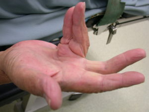
Often, conservative treatment with anti-inflammatory drugs and immobilization of the wrist using a forearm wrist splint can be used (v.a. during the night, so-called. Night splint) achieve a decrease in symptoms. Very advanced nerve damage can often be recognized by the muscle weakening of the ball of the thumb, the picture shows a dent on the ball of the thumb of the left hand. This is the visible correlate of the decrease in strength, which many patients complain about.

If this does not lead to a sufficient improvement in the complaint, the median nerve can be relieved by a small operation. The principle of operation consists in this, to cut through the firm connective tissue covering the nerve and flexor tendons in the area of the carpal tunnel and thereby relieve the nerves. A neurophysiological examination by a neurologist before the operation is essential to confirm the diagnosis and correctly assess the indication for surgery.
The operation to relieve the median nerve can be performed openly or endoscopically. Both OP- The results of procedures are comparable and usually lead to freedom from symptoms. In the so-called. open surgery is performed over an approx. 3 cm long skin incision in the area of the palm-side wrist of the nerve exposed under sight, after all constricting structures have been completely removed, only the skin is closed again.
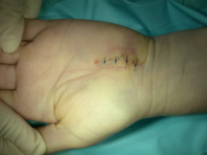
In the endoscopic procedure, an approx. 2 cm long transverse skin incision in the flexor crease of the wrist, an endoscopic knife is inserted under the constricting ligament in the wrist and the ligament is severed in sight. The advantage of the endoscopic procedure is a somewhat faster restoration of resilience.

However, not all patients are suitable for this procedure. An individual decision is made in each case in a personal conversation.
The chances of complete resolution of symptoms are all the better, the less severe the nerve damage before the operation. In the case of advanced nerve damage, the recovery of the nerve and thus the return of feeling can take more than a year or remain incomplete, but even then there is usually a clear improvement in the symptoms, so that the operation is recommended even with a complete loss of sensitivity.
The occupation can in general after 2-6 Weeks (wide range depending on the load) to be resumed.
Rhizarthrosis is the most common arthritis in the hand and affects 70-90% the population. It is arthrosis of the thumb saddle joint, that is, between the metacarpal bone of the thumb and the large polygonal bone (x in the X-ray image below). Usually the joint is swollen (Fig.) and hurts, often the pain cannot be precisely localized to the joint, the symptoms often affect the whole thumb and often radiate to the forearm.

The joint space is on the x-ray (arrow) canceled, the metacarpal has already shifted.

As long as pain only occurs under particular stress and subsides after a short recovery period, no intervention is required. Conservative treatment methods such as. a supporting saddle joint orthosis, anti-inflammatory medications or poultices can help here. Injections or X-ray stimulation can also be temporary, however, it does not provide permanent relief. However, if the pain occurs on a daily basis and interferes with almost all activities in daily life, an operation makes sense. Depending on age, Work demands and expectations as well as the severity of osteoarthritis on the saddle joint and on the neighboring joints are used in various surgical methods. The most frequently used surgical procedure is the so-called Resection arthroplasty. This means the removal of the large polygonal bone and the filling of the resulting gap with tendon tissue from the environment. The drawing shows the OP technology schematically, the filled space appears empty in the X-ray image, because the soft tissue is not radiopaque.

Scheme drawing of the resection arthroplasty: FCR: Flexor carpi radialis (Wrist flexion on the spokes side), APL: Abduktor thumb (Long thumb abductor tendon) and x-ray.

Because the so-called APL tendon is used as a replacement for the removed carpal bone (long thumb abductor tendon) is used, The operation is also called APL plastic. The advantage of this procedure is the simple operating principle, the preservation of the full mobility of the thumb and the extensive absence of complications. In everyday life there is usually extensive freedom from symptoms. A minor disadvantage is a certain loss of strength, when gripping firmly or when there is constant need for a firm grip e.g.. noticeable during professional activity.
Therefore, the process is not suitable for people who work in a manual or otherwise difficult manner. For this group of patients, the Arthrodese, thus a stiffening of the saddle joint. Thereby the two bones, between which there is painful osteoarthritis, firmly connected, so that they form a unit, so to speak. Then the pain will also go away. The advantage of the firm, pain-free grip comes at the expense of a certain restriction of movement, which is hardly noticeable in everyday life, and the additional low risk compared to resection arthroplasty, that the bony connection between the two bones does not occur. This would require revision surgery, but it rarely happens.


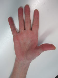
The images show the x-ray after arthrodesis of the saddle joint and illustrate the functional result with good preservation of the mobility of the operated thumb, which is by far sufficient for everyday activities.
Other possible surgical procedures are ligament surgery for early osteoarthritis in patients under 40 Years, which is rarely used. Artificial joint replacements and the implantation of placeholders have so far not found their way into routine hand surgery due to high complication rates and should primarily be reserved for use within scientific studies.
Which surgical procedure is most suitable in each individual case, should be discussed in an individual interview.
Tendon severance caused by an injury naturally requires immediate surgical treatment, the injured person should immediately seek surgical treatment. The variety of injury patterns is too great, to be shown in detail here.
Spontaneous tendon ruptures occur most frequently on the extensor mechanisms of the long finger end joints (‚Mallet-Finger‘) and at the thumb joint (Long extensor tendon ruptured) on.
Mallet Finger
The cause is usually a banal toasting a finger, which leads to an acute stretch deficit. In the acute phase there is usually no pain worth mentioning. The treatment is conservative by consistently immobilizing the end joint in a special finger splint (so-called. Stack’s rail) For 8 Weeks without a break. The duration of treatment can often extend to several months if unsuccessful. Complete healing with restoration of the original condition before the injury is rare, however, a stretching function can usually be restored, which is functionally and cosmetically acceptable. If the stretching deficit persists, surgery can also be used. There are different surgical methods, none is really reliable, therefore, the operation is only recommended in very stubborn cases.
Long extensor tendon ruptured (Extensor EPL Sehnt)
The rupture of the EPL tendon leads to an acute extension deficit of the thumb, this is a clear handicap, therefore surgical treatment is always indicated. The cause of the rupture is usually spontaneous degeneration and weakening of the tendon, which ultimately leads to a rupture with loss of extension of the thumb for no particular reason. The affected patient usually only feels a brief, slight pain on the spinal side of the wrist. Suturing the tendon is usually not possible, the function of the ruptured tendon is restored by repositioning the extensor tendon of the index finger (Extensor indicis Sehne) on the EPL tendon, relearning the function usually takes place in the context of physiotherapy without any problems. The extension function of the index finger does not suffer from this measure, because the index finger has another extensor tendon (so-called. Extensor digitorum Sehnt)
will follow shortly…